| Description |
The Human leukocyte antigen G (HLA-G) ELISA Kit is specifically designed to standardize the detection of HLA-G in serum, plasma, or tissue homogenates. It helps to streamline the experiment and obtain ideal results due to its excellent performances in specificity, sensitivity, precision, linearity, recovery, and lot-to-lot consistency. The assay combines the sandwich-ELISA mechanism with enzyme-substrate chromogenic reaction to complete the detection. The solution color develops in proportion to the HLA-G concentrations in the sample. And the color intensity can be measured at 450 nm via a microplate reader. HLA-G is a non-classical HLA class I antigen with diverse protein isoforms. HLA-G acts as an immune-modulatory molecule and plays an important role in fetal-maternal immune tolerance during pregnancy by interacting with immune cells of both innate and adaptive responses. It is restricted to several healthy adult tissues, including the extravillous cytotrophoblasts, thymic medulla, ... Read more |
| Target Name |
major histocompatibility complex, class I, G |
| Alternative Names |
B2 microglobulin ELISA Kit; DADB-15K14.8 ELISA Kit; HLA 6.0 ELISA Kit; HLA class I histocompatibility antigen alpha chain G ELISA Kit; HLA class I histocompatibility antigen; alpha chain G ELISA Kit; HLA class I molecule ELISA Kit; HLA G ELISA Kit; HLA G antigen ELISA Kit; HLA G histocompatibility antigen class I G ELISA Kit; HLA G3 ELISA Kit; HLA-G ELISA Kit; HLA-G histocompatibility antigen; class I ELISA Kit; HLA60 ELISA Kit; HLAG ELISA Kit; HLAG_HUMAN ELISA Kit; Major histocompatibility complex class I G ELISA Kit; MHC class I antigen ELISA Kit; MHC class I antigen G ELISA Kit; MHC G ELISA Kit; T-cell A locus ELISA Kit; TCA ELISA Kit |
| Abbreviation |
HLA-G |
| Species |
Homo sapiens (Human) |
| Sample Types |
serum, plasma, tissue homogenates |
| Detection Range |
1.25 ng/mL-80 ng/mL |
| Sensitivity |
0.31 ng/mL |
| Assay Time |
1-5h |
| Sample Volume |
50-100ul |
| Detection Wavelength |
450 nm |
| Research Area |
Immunology |
| Assay Principle |
quantitative |
| Measurement |
Sandwich |
| Precision |
| Intra-assay Precision (Precision within an assay): CV% |
| Three samples of known concentration were tested twenty times on one plate to assess. |
|
| Inter-assay Precision (Precision between assays): CV% |
| Three samples of known concentration were tested in twenty assays to assess. |
|
|
| |
|
|
|
|
|
|
|
| Linearity |
| To assess the linearity of the assay, samples were spiked with high concentrations of human HLA-G in various matrices and diluted with the Sample Diluent to produce samples with values within the dynamic range of the assay. |
| |
Sample |
Serum(n=4) |
|
| 1:1 |
Average % |
93 |
|
| Range % |
86-98 |
|
| 1:2 |
Average % |
103 |
|
| Range % |
97-108 |
|
| 1:4 |
Average % |
93 |
|
| Range % |
85-98 |
|
| 1:8 |
Average % |
97 |
|
| Range % |
91-101 |
|
|
| Recovery |
| The recovery of human HLA-G spiked to levels throughout the range of the assay in various matrices was evaluated. Samples were diluted prior to assay as directed in the Sample Preparation section. |
| Sample Type |
Average % Recovery |
Range |
|
| Serum (n=5) |
86 |
80-94 |
|
| EDTA plasma (n=4) |
96 |
92-104 |
|
| |
|
|
|
|
|
|
| |
|
|
|
|
|
|
|
| Typical Data |
| These standard curves are provided for demonstration only. A standard curve should be generated for each set of samples assayed. |
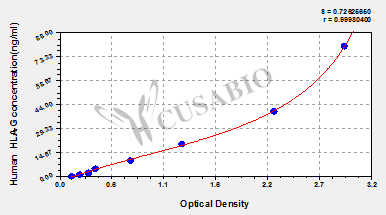
| ng/ml |
OD1 |
OD2 |
Average |
Corrected |
|
| 80 |
2.961 |
2.960 |
2.961 |
2.828 |
|
| 40 |
2.252 |
2.205 |
2.229 |
2.096 |
|
| 20 |
1.266 |
1.286 |
1.276 |
1.143 |
|
| 10 |
0.731 |
0.755 |
0.743 |
0.610 |
|
| 5 |
0.368 |
0.400 |
0.384 |
0.251 |
|
| 2.5 |
0.302 |
0.322 |
0.312 |
0.179 |
|
| 1.25 |
0.227 |
0.215 |
0.221 |
0.088 |
|
| 0 |
0.134 |
0.132 |
0.133 |
|
|
|
|
| Materials provided |
- A micro ELISA plate ---The 96-well plate has been pre-coated with an anti-human HLA-G antibody. This dismountable microplate can be divided into 12 x 8 strip plates.
- Two vials lyophilized standard ---Dilute a bottle of the standard at dilution series, read the OD values, and then draw a standard curve.
- One vial Biotin-labeled HLA-G antibody (100 x concentrate) (120 μl/bottle) ---Act as the detection antibody.
- One vial HRP-avidin (100 x concentrate) (120 μl/bottle) ---Bind to the detection antibody and react with the TMB substrate to make the solution chromogenic.
- One vial Biotin-antibody Diluent (15 ml/bottle) ---Dilute the Biotin-antibody.
- One vial HRP-avidin Diluent (15 ml/bottle) ---Dilute the HRP-avidin solution.
- One vial Sample Diluent (50 ml/bottle)---Dilute the sample to an appropriate concentration.
- One vial Wash Buffer (25 x concentrate) (20 ml/bottle) ---Wash away unbound or free substances.
- One vial TMB Substrate (10 ml/bottle) ---Act as the chromogenic agent. TMB interacts with HRP, eliciting the solution turns blue.
- One vial Stop Solution (10 ml/bottle) ---Stop the color reaction. The solution color immediately turns from blue to yellow.
- Four Adhesive Strips (For 96 wells) --- Cover the microplate when incubation.
- An instruction manual
|
| Materials not provided |
- A microplate reader capable of measuring absorbance at 450 nm, with the correction wavelength set at 540 nm or 570 nm.
- An incubator can provide stable incubation conditions up to 37°C±5°C.
- Centrifuge
- Vortex
- Squirt bottle, manifold dispenser, or automated microplate washer
- Absorbent paper for blotting the microtiter plate
- 50-300ul multi-channel micropipette
- Pipette tips
- Single-channel micropipette with different ranges
- 100ml and 500ml graduated cylinders
- Deionized or distilled water
- Timer
- Test tubes for dilution
|
| Storage |
Store at 2-8°C. Please refer to protocol. |
| Lead Time |
3-5 working days |