Human Vitamin D-binding protein,DBP ELISA Kit
Product Details
>
| Description |
The Human Vitamin D-binding protein (DBP) ELISA Kit is used to detect and quantify the concentrations of DBP in serum, plasma, and tissue homogenates. This kit exclusively recognizes human DBP protein. It adopts the Competitive ELISA technique in which HRP-conjugated DBP and DBP in samples or standards compete for binding to the pre-coated DBP. And the chromogenic reaction is triggered after the addition of TMB substrate solution. After adding the stop solution, the color development is immediately terminated and the color turns from blue to yellow. The intensity of the color is negatively relevant to the levels of DBP in the sample. This ELISA kit has been confirmed to have high sensitivity, excellent specificity, premium precision, high recovery, and lot-to-lot consistency. See the product instructions for more details. DBP, also known as Group-specific Component (GC), is the key transport protein for vitamin D metabolites circulating in the blood. It is primarily produced by hepatic... Read more |
| Target Name |
group-specific component (vitamin D binding protein) |
| Alternative Names |
DBP ELISA Kit; DBP/GC ELISA Kit; GC ELISA Kit; Gc globulin ELISA Kit; Gc-globulin ELISA Kit; GRD3 ELISA Kit; Group specific component ELISA Kit; Group specific component vitamin D binding protein ELISA Kit; Group-specific component ELISA Kit; hDBP ELISA Kit; VDB ELISA Kit; VDBG ELISA Kit; VDBP ELISA Kit; Vitamin D binding alpha globulin ELISA Kit; Vitamin D-binding protein ELISA Kit; VTDB_HUMAN ELISA Kit |
| Abbreviation |
GC |
| Species |
Homo sapiens (Human) |
| Sample Types |
serum, plasma, tissue homogenates |
| Detection Range |
0.156 μg/mL-10 μg/mL |
| Sensitivity |
0.039 μg/mL |
| Assay Time |
1-5h |
| Sample Volume |
50-100ul |
| Detection Wavelength |
450 nm |
| Research Area |
Signal Transduction |
| Assay Principle |
quantitative |
| Measurement |
Competitive |
| Precision |
| Intra-assay Precision (Precision within an assay): CV% |
| Three samples of known concentration were tested twenty times on one plate to assess. |
|
| Inter-assay Precision (Precision between assays): CV% |
| Three samples of known concentration were tested in twenty assays to assess. |
|
|
| |
|
|
|
|
|
|
|
| Linearity |
| To assess the linearity of the assay, samples were spiked with high concentrations of human DBP in various matrices and diluted with the Sample Diluent to produce samples with values within the dynamic range of the assay. |
| |
Sample |
Serum(n=4) |
|
| 1:20 |
Average % |
87 |
|
| Range % |
82-93 |
|
| 1:40 |
Average % |
94 |
|
| Range % |
89-99 |
|
| 1:80 |
Average % |
92 |
|
| Range % |
87-96 |
|
| 1:160 |
Average % |
95 |
|
| Range % |
90-100 |
|
|
| Recovery |
| The recovery of human DBP spiked to levels throughout the range of the assay in various matrices was evaluated. Samples were diluted prior to assay as directed in the Sample Preparation section. |
| Sample Type |
Average % Recovery |
Range |
|
| Serum (n=5) |
92 |
85-97 |
|
| EDTA plasma (n=4) |
96 |
90-101 |
|
| |
|
|
|
|
|
|
| |
|
|
|
|
|
|
|
| Typical Data |
| These standard curves are provided for demonstration only. A standard curve should be generated for each set of samples assayed. |
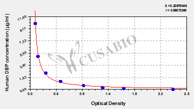
| μg/ml |
OD1 |
OD2 |
Average |
|
|
| 10 |
0.102 |
0.104 |
0.103 |
|
|
| 5 |
0.155 |
0.151 |
0.153 |
|
|
| 2.5 |
0.282 |
0.293 |
0.288 |
|
|
| 1.25 |
0.548 |
0.541 |
0.545 |
|
|
| 0.625 |
0.985 |
0.923 |
0.954 |
|
|
| 0.312 |
1.328 |
1.256 |
1.292 |
|
|
| 0.156 |
1.725 |
1.598 |
1.662 |
|
|
| 0 |
2.614 |
2.444 |
2.529 |
|
|
|
|
| Materials provided |
- A 96-well Assay plate --The 96-well plate has been pre-coated with human DBP.
- Standard(Freeze-dried) (1 x 200 μl) --Dilute the standard at dilution series, read the OD values, and then draw a standard curve.
- HRP-conjugated DBP antibody(100 x concentrate) (1 x 60 μl) --Bind to the DBP, and HRP catalyzes the TMB to elicit a chromogenic reaction.
- HRP-conjugate Diluent (1 x 10 ml) --Dilute the HRP-conjugated DBP antibody solution.
- Sample Diluent (2 x 20 ml) --Reconstitute the standard and dilute the sample to an appropriate concentration.
- Wash Buffer (25x concentrate) (1 x 20 ml) --Wash away unbound or free substances.
- TMB Substrate (1x 10 ml) --Act as the chromogenic agent. TMB interacts with HRP, eliciting the solution turns blue.
- Stop Solution (1 x 10ml) --Stop the color reaction. The solution color immediately turns from blue to yellow.
- Four Adhesive Strips (For 96 wells)
- An Instruction manual
|
| Materials not provided |
- A microplate reader capable of measuring absorbance at 450 nm, with the correction wavelength set at 540 nm - 570 nm.
- An incubator that can provide stable incubation conditions up to 37°C±5°C.
- Centrifuge
- Vortex
- Squirt bottle, manifold dispenser, or automated microplate washer
- Absorbent paper for blotting the microtiter plate
- 50-300ul multi-channel micropipette
- Pipette tips
- Single-channel micropipette with different ranges
- 100ml and 500ml graduated cylinders
- Deionized or distilled water
- Timer
- Test tubes for dilution
|
| Storage |
Store at 2-8°C. Please refer to protocol. |
| Lead Time |
3-5 working days |