Mouse C-Peptide ELISA Kit
Product Details
| Description |
The Mouse C-peptide ELISA Kit is used to quantitatively measure the concentrations of C-peptide in mouse serum and plasma. This assay employs the quantitative sandwich enzyme immunoassay technique, in which C-peptide in the samples or standards are sandwiched between pre-coated C-peptide antibody and HRP-conjugated antibody specific for C-peptide. Following a wash to remove any unbound reagent, the TMB substrate solution is added to the wells and color develops in proportion to the amount of C-peptide bound in the initial step. The color development is stopped and the intensity of the color is measured at 450 nm via a microplate reader. This kit has been validated with various criteria, including sensitivity, specificity, precision (less than 15%), linearity, and recovery. The product instructions are access to more information.
C-peptide is a 31-amino-acid-peptide released from the pancreatic beta cells during cleavage of insulin from proinsulin. It does not actually affect the blood sugar. C-peptide is produced in equimolar amounts to endogenous insulin, so it is useful as a marker of insulin production. Moreover, C-peptide degrades more slowly in the body than insulin does. It is therefore a useful and widely used method of assessing pancreatic beta-cell function. The C-peptide test is used to help evaluate blood sugar disorders, including type 1 or type 2 diabetes. A normal C-peptide range is 0.5 to 2.0 nanograms per milliliter. Most C-peptide is metabolized by the kidneys with 5-10% then excreted unaltered in the urine, which makes it inaccurate in the measurement of C-peptide in individuals with chronic kidney disease.
|
| Target Name |
C-Peptide |
| Abbreviation |
C-Peptide |
| Species |
Mus musculus (Mouse) |
| Sample Types |
serum, plasma |
| Detection Range |
2.86 ng/mL-50 ng/mL |
| Sensitivity |
1.786 ng/mL |
| Assay Time |
1-5h |
| Sample Volume |
50-100ul |
| Detection Wavelength |
450 nm |
| Research Area |
Metabolism |
| Assay Principle |
quantitative |
| Measurement |
Sandwich |
| Precision |
| Intra-assay Precision (Precision within an assay): CV% |
| Three samples of known concentration were tested twenty times on one plate to assess. |
|
| Inter-assay Precision (Precision between assays): CV% |
| Three samples of known concentration were tested in twenty assays to assess. |
|
|
| |
|
|
|
|
|
|
|
| Linearity |
| To assess the linearity of the assay, samples were spiked with high concentrations of mouse C-Peptide in various matrices and diluted with the Sample Diluent to produce samples with values within the dynamic range of the assay. |
| |
Sample |
Serum(n=4) |
|
| 1:1 |
Average % |
93 |
|
| Range % |
85-99 |
|
| 1:2 |
Average % |
90 |
|
| Range % |
85-96 |
|
| 1:4 |
Average % |
95 |
|
| Range % |
87-106 |
|
| 1:8 |
Average % |
95 |
|
| Range % |
89-100 |
|
|
| Recovery |
| The recovery of mouse C-Peptide spiked to levels throughout the range of the assay in various matrices was evaluated. Samples were diluted prior to assay as directed in the Sample Preparation section. |
| Sample Type |
Average % Recovery |
Range |
|
| Serum (n=5) |
96 |
89-100 |
|
| EDTA plasma (n=4) |
95 |
91-103 |
|
| |
|
|
|
|
|
|
| |
|
|
|
|
|
|
|
| Typical Data |
| These standard curves are provided for demonstration only. A standard curve should be generated for each set of samples assayed. |
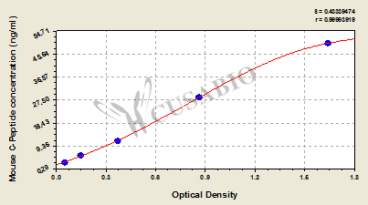
| ng/ml |
OD1 |
OD2 |
Average |
|
|
| 2.86 |
0.055 |
0.059 |
0.057 |
|
|
| 5.71 |
0.147 |
0.154 |
0.151 |
|
|
| 11.43 |
0.367 |
0.372 |
0.370 |
|
|
| 28.57 |
0.846 |
0.855 |
0.851 |
|
|
| 50 |
1.618 |
1.607 |
1.613 |
|
|
|
|
| Materials provided |
- A micro ELISA plate ---The 96-well plate has been pre-coated with an anti-mouse C-peptide antibody.
- Six vials lyophilized standard ---Dilute a bottle of the standard at dilution series, read the OD values, and then draw a standard curve.
- One vial HRP-conjugated C-peptide antibody(6 ml/bottle) ---Bind to the C-peptide in the samples or standards and react with the substrate to make the solution chromogenic.
- One vial Wash Buffer (20x concentrate) (15ml/bottle) ---Wash away unbound or free substances.
- Substrate A (1 x 7 ml)
- Substrate B (1 x 7 ml)
- One vial Stop Solution (7ml/bottle) ---Stop the color reaction. The solution color immediately turns from blue to yellow.
- Four Adhesive Strips (For 96 wells) ---Cover the microplate when incubation.
- An instruction manual
|
| Materials not provided |
- A microplate reader capable of measuring absorbance at 450 nm, with the correction wavelength set at 600 nm or 630 nm.
- An incubator can provide stable incubation conditions up to 37°C±5°C.
- Centrifuge
- Vortex
- Squirt bottle, manifold dispenser, or automated microplate washer
- Absorbent paper for blotting the microtiter plate
- 50-300ul multi-channel micropipette
- Pipette tips
- Single-channel micropipette with different ranges
- 100ml and 500ml graduated cylinders
- Deionized or distilled water
- Timer
- Test tubes for dilution
|
| Storage |
Store at 2-8°C. Please refer to protocol. |
| Lead Time |
3-5 working days |