| Description |
This Mouse C-Reactive Protein (CRP) ELISA Kit is used to quantitatively determine mouse CRP concentrations in serum, plasma, cell culture supernates, or tissue homogenates. CRP is an acute inflammatory protein that exhibits significantly elevated [removed]1,000-fold) at sites of infection or inflammation. It has been traditionally used as a marker of infection and cardiovascular events. Increasing evidence shows that CRP plays important role in inflammatory processes and host defense to infection including the complement pathway, apoptosis, phagocytosis, NO release, and the production of cytokines, particularly IL-6 and TNF-α.
This kit employs the quantitative sandwich enzyme immunoassay technique to measure the levels of mouse CRP in the sample. Antibody specific for CRP has been pre-coated onto the microplate. Standards and samples are pipetted into the wells and any CRP present is bound by the immobilized antibody. After removing any unbound substances, a biotin-conjugated CRP antibody is added to the wells. After washing, avidin conjugated HRP is added to the wells, forming an antibody-antigen-enzyme-labeled antibody complex. Following a wash to remove any unbound HRP-avidin, the TMB substrate solution is added to the wells, and color develops into blue. The color changes from blue to yellow after the addition of stop solution into the wells. The intensity of the color is in proportion to the amount of CRP bound in the initial step. The absorbance (OD value) was determined by the microplate reader at the wavelength of 450nm, and the concentration of mouse NT-proBNP was calculated through the standard curve. Each manufactured lot of this ELISA kit is quality tested for criteria such as sensitivity, specificity, precision, linearity, recovery, and lot-to-lot consistency. See the instructions for more information on validation
|
| Target Name |
C-reactive protein, pentraxin-related |
| Alternative Names |
Crp ELISA Kit; Ptx1C-reactive protein ELISA Kit |
| Abbreviation |
CRP |
| Species |
Mus musculus (Mouse) |
| Sample Types |
serum, plasma, cell culture supernates, tissue homogenates |
| Detection Range |
15.6 ng/mL-1000 ng/mL |
| Sensitivity |
3.9 ng/mL |
| Assay Time |
1-5h |
| Sample Volume |
50-100ul |
| Detection Wavelength |
450 nm |
| Research Area |
Immunology |
| Assay Principle |
quantitative |
| Measurement |
Sandwich |
| Precision |
| Intra-assay Precision (Precision within an assay): CV% |
| Three samples of known concentration were tested twenty times on one plate to assess. |
|
| Inter-assay Precision (Precision between assays): CV% |
| Three samples of known concentration were tested in twenty assays to assess. |
|
|
| |
|
|
|
|
|
|
|
| Linearity |
| To assess the linearity of the assay, samples were spiked with high concentrations of mouse CRP in various matrices and diluted with the Sample Diluent to produce samples with values within the dynamic range of the assay. |
| |
Sample |
Serum(n=4) |
|
| 1:20 |
Average % |
90 |
|
| Range % |
86-93 |
|
| 1:40 |
Average % |
103 |
|
| Range % |
99-106 |
|
| 1:80 |
Average % |
93 |
|
| Range % |
87-96 |
|
| 1:160 |
Average % |
98 |
|
| Range % |
93-101 |
|
|
| Recovery |
| The recovery of mouse CRP spiked to levels throughout the range of the assay in various matrices was evaluated. Samples were diluted prior to assay as directed in the Sample Preparation section. |
| Sample Type |
Average % Recovery |
Range |
|
| Serum (n=5) |
94 |
89-97 |
|
| EDTA plasma (n=4) |
97 |
90-100 |
|
| |
|
|
|
|
|
|
| |
|
|
|
|
|
|
|
| Typical Data |
| These standard curves are provided for demonstration only. A standard curve should be generated for each set of samples assayed. |
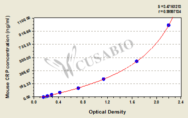
| ng/ml |
OD1 |
OD2 |
Average |
Corrected |
|
| 1000 |
2.206 |
2.124 |
2.165 |
1.995 |
|
| 500 |
1.661 |
1.655 |
1.658 |
1.488 |
|
| 250 |
1.138 |
1.122 |
1.130 |
0.960 |
|
| 125 |
0.727 |
0.721 |
0.724 |
0.554 |
|
| 62.5 |
0.424 |
0.435 |
0.430 |
0.260 |
|
| 31.2 |
0.292 |
0.305 |
0.299 |
0.129 |
|
| 15.6 |
0.228 |
0.234 |
0.231 |
0.061 |
|
| 0 |
0.168 |
0.172 |
0.170 |
|
|
|
|
| Materials provided |
- An assay plate --- The 96-well plate is composed of 12 x 8 strip plates and can be taken apart.
- Two bottles of lyophilized standard --- Reconstitute one bottle of the standard with sample diluent at dilution series and draw the standard curve.
- Biotin-labeled CRPantibody (100 x concentrate) 1 x 120 μl --- Act as the detection antibody and need to be diluted before use.
- HRP-avidin (100 x concentrate) 1 x 120 μl --- Catalyze the TMB substrate for color development and need to be diluted before use.
- Biotin-antibody Diluent 1 x 15 ml --- Dilute the Biotin-antibody.
- HRP-avidin Diluent 1 x 15 ml --- Dilute the HRP-avidin.
- Sample Diluent 1 x 50 ml --- Reconstitute the standard and dilute the sample to an appropriate concentration.
- Wash Buffer (25 x concentrate) 1 x 20 ml --- Wash away the unbound solution and non-specific substances.
- TMB Substrate 1 x 10 ml --- Act as the chromogenic agent. TMB interacts with HRP, eliciting the solution turns blue.
- Stop Solution 1 x 10 ml --- Add stop solution and stop the color development. The solution color immediately turns from blue to yellow.
- Four Adhesive Strips (For 96 wells) --- Seal the microtiter plate when incubation.
- An instruction manual
|
| Materials not provided |
- A microplate reader capable of measuring absorbance at 450 nm, with the correction wavelength set at 540 nm or 570 nm.
- An incubator can provide stable incubation conditions up to 37°C±5°C.
- Centrifuge
- Vortex
- Squirt bottle, manifold dispenser, or automated microplate washer
- Absorbent paper for blotting the microtiter plate
- 50-300ul multi-channel micropipette
- Pipette tips
- Single-channel micropipette with different ranges
- 100ml and 500ml graduated cylinders
- Deionized or distilled water
- Timer
- Test tubes for dilution
|
| Storage |
Store at 2-8°C. Please refer to protocol. |
| Lead Time |
3-5 working days |