| Description |
The Mouse cyclooxygenase-2 (COX-2) ELISA Kit is designed to measure COX-2 in biological samples, including serum, plasma, tissue homogenates, or cell lysates, quantitatively. This kit has been verified by multiple tests with advantages of high sensitivity, strong specificity, precision less than 10%, and lot-to-lot consistency. Based on the Sandwich-ELISA technique in combination with the enzyme-substrate chromogenic reaction as well as colorimetric measurement, the levels of COX-2 in the sample can be calculated.
COX-2, also named PTGS2, is an inducible COX isoform in normal tissue such as colorectal, kidney, reproductive organs, and stomach. However, COX2 is continuously up-regulated during both inflammation and some types of cancer, such as adenocarcinoma, cholangiocarcinoma, and hepatocellular carcinoma. It regulates and mediates the biosynthesis and release of prostaglandins using arachidonic acid (AA) as the substrate. COX-2 and its product prostaglandin E2 (PGE2) enhances cell proliferation, cell survival, and tumor invasion mainly in colorectal, breast, and prostate cancers, as well as hematological malignancies, thus facilitating tumor progression.
|
| Target Name |
prostaglandin-endoperoxide synthase 2 (prostaglandin G/H synthase and cyclooxygenase) |
| Alternative Names |
Ptgs2 ELISA Kit; Cox-2 ELISA Kit; Cox2 ELISA Kit; Pghs-b ELISA Kit; Tis10 ELISA Kit; Prostaglandin G/H synthase 2 ELISA Kit; EC 1.14.99.1 ELISA Kit; Cyclooxygenase-2 ELISA Kit; COX-2 ELISA Kit; Glucocorticoid-regulated inflammatory cyclooxygenase ELISA Kit; Gripghs ELISA Kit; Macrophage activation-associated marker protein P71/73 ELISA Kit; PES-2 ELISA Kit; PHS II ELISA Kit; Prostaglandin H2 synthase 2 ELISA Kit; PGH synthase 2 ELISA Kit; PGHS-2 ELISA Kit; Prostaglandin-endoperoxide synthase 2 ELISA Kit; TIS10 protein ELISA Kit |
| Abbreviation |
PTGS2 |
| Species |
Mus musculus (Mouse) |
| Sample Types |
serum, plasma, tissue homogenates, cell lysates |
| Detection Range |
31.25 pg/mL-2000 pg/mL |
| Sensitivity |
7.8 pg/mL |
| Assay Time |
1-5h |
| Sample Volume |
50-100ul |
| Detection Wavelength |
450 nm |
| Research Area |
Metabolism |
| Assay Principle |
quantitative |
| Measurement |
Sandwich |
| Precision |
| Intra-assay Precision (Precision within an assay): CV% |
| Three samples of known concentration were tested twenty times on one plate to assess. |
|
| Inter-assay Precision (Precision between assays): CV% |
| Three samples of known concentration were tested in twenty assays to assess. |
|
|
| |
|
|
|
|
|
|
|
| Linearity |
| To assess the linearity of the assay, samples were spiked with high concentrations of mouse COX-2 in various matrices and diluted with the Sample Diluent to produce samples with values within the dynamic range of the assay. |
| |
Sample |
Serum(n=4) |
|
| 1:1 |
Average % |
88 |
|
| Range % |
80-92 |
|
| 1:2 |
Average % |
98 |
|
| Range % |
91-105 |
|
| 1:4 |
Average % |
100 |
|
| Range % |
92-110 |
|
| 1:8 |
Average % |
93 |
|
| Range % |
86-98 |
|
|
| Recovery |
| The recovery of mouse COX-2 spiked to levels throughout the range of the assay in various matrices was evaluated. Samples were diluted prior to assay as directed in the Sample Preparation section. |
| Sample Type |
Average % Recovery |
Range |
|
| Serum (n=5) |
96 |
89-98 |
|
| EDTA plasma (n=4) |
96 |
90-100 |
|
| |
|
|
|
|
|
|
| |
|
|
|
|
|
|
|
| Typical Data |
| These standard curves are provided for demonstration only. A standard curve should be generated for each set of samples assayed. |
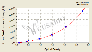
| pg/ml |
OD1 |
OD2 |
Average |
Corrected |
|
| 2000 |
2.660 |
2.654 |
2.657 |
2.547 |
|
| 1000 |
2.107 |
2.046 |
1.994 |
1.884 |
|
| 500 |
1.494 |
1.385 |
1.440 |
1.330 |
|
| 250 |
0.932 |
0.911 |
0.922 |
0.812 |
|
| 125 |
0.502 |
0.489 |
0.496 |
0.386 |
|
| 62.5 |
0.403 |
0.398 |
0.415 |
0.305 |
|
| 31.25 |
0.223 |
0.207 |
0.215 |
0.105 |
|
| 0 |
0.114 |
0.106 |
0.110 |
|
|
|
|
| Materials provided |
- A micro ELISA plate ---The 96-well plate has been pre-coated with an anti-mouse COX-2 antibody. This dismountable microplate can be divided into 12 x 8 strip plates.
- Two vials lyophilized standard ---Dilute a bottle of the standard at dilution series, read the OD values, and then draw a standard curve.
- One vial Biotin-labeled COX-2 antibody (100 x concentrate) (120 μl/bottle) ---Act as the detection antibody.
- One vial HRP-avidin (100 x concentrate) (120 μl/bottle) ---Bind to the detection antibody and react with the TMB substrate to make the solution chromogenic.
- One vial Biotin-antibody Diluent (15 ml/bottle) ---Dilute the Biotin-antibody.
- One vial HRP-avidin Diluent (15 ml/bottle) ---Dilute the HRP-avidin solution.
- One vial Sample Diluent (50 ml/bottle)---Dilute the sample to an appropriate concentration.
- One vial Wash Buffer (25 x concentrate) (20 ml/bottle) ---Wash away unbound or free substances.
- One vial TMB Substrate (10 ml/bottle) ---Act as the chromogenic agent. TMB interacts with HRP, eliciting the solution turns blue.
- One vial Stop Solution (10 ml/bottle) ---Stop the color reaction. The solution color immediately turns from blue to yellow.
- Four Adhesive Strips (For 96 wells) --- Cover the microplate when incubation.
- An instruction manual
|
| Materials not provided |
- A microplate reader capable of measuring absorbance at 450 nm, with the correction wavelength set at 540 nm or 570 nm.
- An incubator can provide stable incubation conditions up to 37°C±5°C.
- Centrifuge
- Vortex
- Squirt bottle, manifold dispenser, or automated microplate washer
- Absorbent paper for blotting the microtiter plate
- 50-300ul multi-channel micropipette
- Pipette tips
- Single-channel micropipette with different ranges
- 100ml and 500ml graduated cylinders
- Deionized or distilled water
- Timer
- Test tubes for dilution
|
| Storage |
Store at 2-8°C. Please refer to protocol. |
| Lead Time |
3-5 working days |