Mouse Interleukin 2,IL-2 ELISA kit
Product Details
| Description |
The Mouse Interleukin 2 (IL-2) ELISA kit is designed for detection and quantification of human IL-2 in serum, plasma, tissue homogenates, or cell culture supernates. It has been validated with high sensitivity, strong specificity, good linearity, precision less than 10%, high recovery, and consistency across batches. Refer to the product instructions for more details.
The measurement of this assay is based on the Sandwich-ELISA technique. The samples and biotin-labeled IL-2 antibody are successively pipetted into the wells pre-coated with anti-mouse IL-2 antibody. The IL-2 is sandwiched between the two antibodies, forming Ag-Ab-Ag immune complex. HRP-avidin is subsequently added to the wells. After thorough washing, the substrate TMB is added for color development. TMB is converted to blue by HRP and yellow by the stop solution. The intensity of the color is positively correlated with the IL-2 content in the samples.
IL-2 is a pleiotropic cytokine produced by activated T cells. It is involved in various biological processes, including the differentiation and survival of CD4+ T helper subsets and CD4+ T regulatory cells, promotion of the cytolytic activity of natural killer (NK) cells and T cells, and augment of immunoglobulin production via activated B cells. IL-2 controls diverse T cell biology by binding with IL-2R. And IL-2/IL-2R signaling is important for T cell activation through the JAK1-STAT5 signaling pathway.
Hide more
|
| Target Name |
interleukin 2 |
| Alternative Names |
Il2 ELISA kit; Il-2Interleukin-2 ELISA kit; IL-2 ELISA kit; T-cell growth factor ELISA kit; TCGF ELISA kit |
| Abbreviation |
IL2 |
| Species |
Mus musculus (Mouse) |
| Sample Types |
serum, plasma, tissue homogenates, cell culture supernates |
| Detection Range |
15.6 pg/mL-1000 pg/mL |
| Sensitivity |
3.9 pg/mL |
| Assay Time |
1-5h |
| Sample Volume |
50-100ul |
| Detection Wavelength |
450 nm |
| Research Area |
Immunology |
| Assay Principle |
quantitative |
| Measurement |
Sandwich |
| Precision |
| Intra-assay Precision (Precision within an assay): CV% |
| Three samples of known concentration were tested twenty times on one plate to assess. |
|
| Inter-assay Precision (Precision between assays): CV% |
| Three samples of known concentration were tested in twenty assays to assess. |
|
|
| |
|
|
|
|
|
|
|
| Linearity |
| To assess the linearity of the assay, samples were spiked with high concentrations of mouse IL-2 in various matrices and diluted with the Sample Diluent to produce samples with values within the dynamic range of the assay. |
| |
Sample |
Serum(n=4) |
|
| 1:1 |
Average % |
87 |
|
| Range % |
81-93 |
|
| 1:2 |
Average % |
99 |
|
| Range % |
93-105 |
|
| 1:4 |
Average % |
95 |
|
| Range % |
90-100 |
|
| 1:8 |
Average % |
96 |
|
| Range % |
90-104 |
|
|
| Recovery |
| The recovery of mouse IL-2 spiked to levels throughout the range of the assay in various matrices was evaluated. Samples were diluted prior to assay as directed in the Sample Preparation section. |
| Sample Type |
Average % Recovery |
Range |
|
| Serum (n=5) |
93 |
88-98 |
|
| EDTA plasma (n=4) |
97 |
90-103 |
|
| |
|
|
|
|
|
|
| |
|
|
|
|
|
|
|
| Typical Data |
| These standard curves are provided for demonstration only. A standard curve should be generated for each set of samples assayed. |
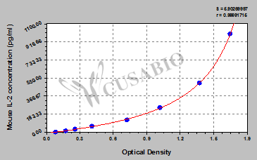
| pg/ml |
OD1 |
OD2 |
Average |
Corrected |
|
| 1000 |
1.729 |
1.652 |
1.691 |
1.597 |
|
| 500 |
1.434 |
1.386 |
1.410 |
1.316 |
|
| 250 |
1.091 |
1.004 |
1.048 |
0.954 |
|
| 125 |
0.765 |
0.729 |
0.747 |
0.653 |
|
| 62.5 |
0.428 |
0.421 |
0.425 |
0.331 |
|
| 31.2 |
0.276 |
0.269 |
0.273 |
0.179 |
|
| 15.6 |
0.193 |
0.179 |
0.186 |
0.092 |
|
| 0 |
0.097 |
0.090 |
0.094 |
|
|
|
|
| Materials provided |
- A micro ELISA plate ---The 96-well plate has been pre-coated with an anti-mouse IL-2 antibody. This dismountable microplate can be divided into 12 x 8 strip plates.
- Two vials lyophilized standard ---Dilute a bottle of the standard at dilution series, read the OD values, and then draw a standard curve.
- One vial Biotin-labeled IL-2 antibody (100 x concentrate) (120 μl/bottle) ---Act as the detection antibody.
- One vial HRP-avidin (100 x concentrate) (120 μl/bottle) ---Bind to the detection antibody and react with the TMB substrate to make the solution chromogenic.
- One vial Biotin-antibody Diluent (15 ml/bottle) ---Dilute the Biotin-antibody.
- One vial HRP-avidin Diluent (15 ml/bottle) ---Dilute the HRP-avidin solution.
- One vial Sample Diluent (50 ml/bottle)---Dilute the sample to an appropriate concentration.
- One vial Wash Buffer (25 x concentrate) (20 ml/bottle) ---Wash away unbound or free substances.
- One vial TMB Substrate (10 ml/bottle) ---Act as the chromogenic agent. TMB interacts with HRP, eliciting the solution turns blue.
- One vial Stop Solution (10 ml/bottle) ---Stop the color reaction. The solution color immediately turns from blue to yellow.
- Four Adhesive Strips (For 96 wells) --- Cover the microplate when incubation.
- An instruction manual
|
| Materials not provided |
- A microplate reader capable of measuring absorbance at 450 nm, with the correction wavelength set at 540 nm or 570 nm.
- An incubator can provide stable incubation conditions up to 37°C±5°C.
- Centrifuge
- Vortex
- Squirt bottle, manifold dispenser, or automated microplate washer
- Absorbent paper for blotting the microtiter plate
- 50-300ul multi-channel micropipette
- Pipette tips
- Single-channel micropipette with different ranges
- 100ml and 500ml graduated cylinders
- Deionized or distilled water
- Timer
- Test tubes for dilution
|
| Storage |
Store at 2-8°C. Please refer to protocol. |
| Lead Time |
3-5 working days |

