| Description |
The Rat growth hormone (GH) ELISA KIT is used to quantitatively measure the levels of CEA in human serum, plasma, cell culture supernates, or tissue homogenates. This assay employs the quantitative sandwich enzyme immunoassay technique, in which GH in the samples or standards are sandwiched between pre-coated GH antibody and Biotin-conjugated antibody specific for GH. The Ab-Ag-Ab complex is labeled by HRP. Following a wash to remove any unbound reagent, the TMB substrate solution is added to the wells and color develops in proportion to the amount of GH bound in the initial step. The color development is stopped and the intensity of the color is measured at 450 nm via a microplate reader. This kit displays many advantages, including sensitivity, specificity, precision, linearity, and recovery. The product instructions are access to more information.
GH is a pituitary gland-produced peptide hormone that spurs growth, cell reproduction, body composition, cell repair, and metabolism in mammalians. The secretion of GH is a part of a negative feedback loop involving IGF-1. Its secretion is stimulated by the growth hormone-releasing hormone (GHRH) and is suppressed by somatostatin. High blood levels of IGF-1 cause a decrease in GH secretion not only directly inhibiting the somatotroph but by stimulating somatostatin release from the hypithalamus. GH deficiency, one of the many causes of dwarfism and short stature, is caused mainly by damage to the hypithalamus or the pituitary gland during fetal development or following birth.
Hide more
|
| Target Name |
growth hormone 1 |
| Alternative Names |
Ghrh ELISA Kit; Somatoliberin ELISA Kit; Growth hormone-releasing factor ELISA Kit; GRF ELISA Kit; Growth hormone-releasing hormone ELISA Kit; GHRH ELISA Kit |
| Abbreviation |
GH1 |
| Species |
Rattus norvegicus (Rat) |
| Sample Types |
serum, plasma, cell culture supernates, tissue homogenates |
| Detection Range |
3.12 pg/mL-200 pg/mL |
| Sensitivity |
0.78 pg/mL |
| Assay Time |
1-5h |
| Sample Volume |
50-100ul |
| Detection Wavelength |
450 nm |
| Research Area |
Signal Transduction |
| Assay Principle |
quantitative |
| Measurement |
Sandwich |
| Precision |
| Intra-assay Precision (Precision within an assay): CV% |
| Three samples of known concentration were tested twenty times on one plate to assess. |
|
| Inter-assay Precision (Precision between assays): CV% |
| Three samples of known concentration were tested in twenty assays to assess. |
|
|
| |
|
|
|
|
|
|
|
| Linearity |
| To assess the linearity of the assay, samples were spiked with high concentrations of rat GH in various matrices and diluted with the Sample Diluent to produce samples with values within the dynamic range of the assay. |
| |
Sample |
Serum(n=4) |
|
| 1:100 |
Average % |
98 |
|
| Range % |
94-105 |
|
| 1:200 |
Average % |
96 |
|
| Range % |
90-102 |
|
| 1:400 |
Average % |
100 |
|
| Range % |
94-106 |
|
| 1:800 |
Average % |
93 |
|
| Range % |
89-97 |
|
|
| Recovery |
| The recovery of rat GH spiked to levels throughout the range of the assay in various matrices was evaluated. Samples were diluted prior to assay as directed in the Sample Preparation section. |
| Sample Type |
Average % Recovery |
Range |
|
| Serum (n=5) |
92 |
87-98 |
|
| EDTA plasma (n=4) |
96 |
91-101 |
|
| |
|
|
|
|
|
|
| |
|
|
|
|
|
|
|
| Typical Data |
| These standard curves are provided for demonstration only. A standard curve should be generated for each set of samples assayed. |
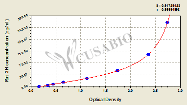
| pg/ml |
OD1 |
OD2 |
Average |
Corrected |
|
| 200 |
2.718 |
2.801 |
2.760 |
2.580 |
|
| 100 |
2.346 |
2.405 |
2.376 |
2.196 |
|
| 50 |
1.754 |
1.763 |
1.759 |
1.579 |
|
| 25 |
1.126 |
1.157 |
1.142 |
0.962 |
|
| 12.5 |
0.657 |
0.668 |
0.663 |
0.483 |
|
| 6.25 |
0.458 |
0.467 |
0.463 |
0.283 |
|
| 3.12 |
0.341 |
0.358 |
0.350 |
0.170 |
|
| 0 |
0.179 |
0.181 |
0.180 |
|
|
|
|
| Materials provided |
- A micro ELISA plate --The 96-well plate has been pre-coated with an anti-rat GH antibody. This dismountable microplate can be divided into 12 x 8 strip plates.
- Two vials lyophilized standard --Dilute a bottle of the standard at dilution series, read the OD values, and then draw a standard curve.
- One vial Biotin-labeled GH antibody (100 x concentrate) (120 μl/bottle) --Act as the detection antibody.
- One vial HRP-avidin (100 x concentrate) (120 μl/bottle) --Bind to the detection antibody and react with the TMB substrate to make the solution chromogenic.
- One vial Biotin-antibody Diluent (15 ml/bottle) --Dilute the high concentration Biotin-antibody to an appropriate working solution.
- One vial HRP-avidin Diluent (15 ml/bottle) --Dilute the high concentration HRP-avidin solution to an appropriate solution.
- One vial Sample Diluent (50 ml/bottle)--Dilute the sample to an appropriate concentration.
- One vial Wash Buffer (25 x concentrate) (20 ml/bottle) --- Wash away unbound or free substances.
- One vial TMB Substrate (10 ml/bottle) --Act as the chromogenic agent. TMB interacts with HRP, eliciting the solution turns blue.
- One vial Stop Solution (10 ml/bottle) --Stop the color reaction. The solution color immediately turns from blue to yellow.
- Four Adhesive Strips (For 96 wells) --Cover the microplate when incubation.
- An instruction manual
|
| Materials not provided |
- A microplate reader capable of measuring absorbance at 450 nm, with the correction wavelength set at 540 nm or 570 nm.
- An incubator can provide stable incubation conditions up to 37°C±5°C.
- Centrifuge
- Vortex
- Squirt bottle, manifold dispenser, or automated microplate washer
- Absorbent paper for blotting the microtiter plate
- 50-300ul multi-channel micropipette
- Pipette tips
- Single-channel micropipette with different ranges
- 100ml and 500ml graduated cylinders
- Deionized or distilled water
- Timer
- Test tubes for dilution
|
| Storage |
Store at 2-8°C. Please refer to protocol. |
| Lead Time |
3-5 working days |