Rat Immunoglobulin E,IgE ELISA Kit
Product Details
| Description |
The product CSB-E07984r is a ready-to-use microwell, strip plate ELISA Kit for quantitative analysis of rat immunoglobulin E(IgE) in serum and plasma. The detection mechanism of this kit is based on the Sandwich-ELISA technique. IgE in the samples or standards is sandwiched between pre-coated IgE antibody and Biotin-conjugated IgE antibody. The IgE/pre-coated IgE antibody/Biotin-conjugated IgE antibody complex is labeled by HRP and then occurs a chromogenic reaction after the addition of the TMB solution. The color intensity is positively related to the IgE in the samples. This kit has undergone rigorous quality tests in multiple parameters, including specificity, sensitivity, precision, linearity, recovery, and lot-to-lot consistency. More details are described in the product instructions.
IgE is a group of antibodies produced by the immune system when overreacting to an allergen. IgE travels to cells including mast cells and basophils and then binds to the Fc receptors on the surface of these cells, inducing these cells to release chemicals and resulting in an allergic reaction. It commonly triggers symptoms in the skin, nose, throat, or lungs. The levels of IgE are normally very low in the blood but get higher when the body is fighting against a parasitic infection or with some immune system conditions. Each type of IgE has a specific "radar" for each type of allergen. That's why some people are only allergic to pollen, while others have allergic reactions to multiple allergens due to more kinds of IgE antibodies present in their bodies.
|
| Target Name |
Immunoglobulin E,IgE |
| Abbreviation |
IgE |
| Species |
Rattus norvegicus (Rat) |
| Sample Types |
serum, plasma |
| Detection Range |
3.12 ng/mL-200 ng/mL |
| Sensitivity |
0.78 ng/mL |
| Assay Time |
1-5h |
| Sample Volume |
50-100ul |
| Detection Wavelength |
450 nm |
| Research Area |
Immunology |
| Assay Principle |
quantitative |
| Measurement |
Sandwich |
| Precision |
| Intra-assay Precision (Precision within an assay): CV% |
| Three samples of known concentration were tested twenty times on one plate to assess. |
|
| Inter-assay Precision (Precision between assays): CV% |
| Three samples of known concentration were tested in twenty assays to assess. |
|
|
| |
|
|
|
|
|
|
|
| Linearity |
| To assess the linearity of the assay, samples were spiked with high concentrations of rat IgE in various matrices and diluted with the Sample Diluent to produce samples with values within the dynamic range of the assay. |
| |
Sample |
Serum(n=4) |
|
| 1:1 |
Average % |
85 |
|
| Range % |
84-93 |
|
| 1:2 |
Average % |
96 |
|
| Range % |
91-103 |
|
| 1:4 |
Average % |
84 |
|
| Range % |
81-89 |
|
| 1:8 |
Average % |
93 |
|
| Range % |
89-97 |
|
|
| Recovery |
| The recovery of rat IgE spiked to levels throughout the range of the assay in various matrices was evaluated. Samples were diluted prior to assay as directed in the Sample Preparation section. |
| Sample Type |
Average % Recovery |
Range |
|
| Serum (n=5) |
92 |
87-98 |
|
| EDTA plasma (n=4) |
95 |
90-99 |
|
| |
|
|
|
|
|
|
| |
|
|
|
|
|
|
|
| Typical Data |
| These standard curves are provided for demonstration only. A standard curve should be generated for each set of samples assayed. |
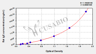
| ng/ml |
OD1 |
OD2 |
Average |
Corrected |
|
| 200 |
2.055 |
2.086 |
2.071 |
1.926 |
|
| 100 |
1.520 |
1.649 |
1.585 |
1.440 |
|
| 50 |
1.101 |
1.156 |
1.129 |
0.984 |
|
| 25 |
0.738 |
0.750 |
0.744 |
0.599 |
|
| 12.5 |
0.417 |
0.445 |
0.431 |
0.286 |
|
| 6.25 |
0.341 |
0.337 |
0.339 |
0.194 |
|
| 3.12 |
0.201 |
0.213 |
0.207 |
0.062 |
|
| 0 |
0.139 |
0.150 |
0.145 |
|
|
|
|
| Materials provided |
- A micro ELISA plate --The 96-well plate has been pre-coated with an anti-rat IgE antibody. This dismountable microplate can be divided into 12 x 8 strip plates.
- Two vials lyophilized standard --Dilute a bottle of the standard at dilution series, read the OD values, and then draw a standard curve.
- One vial Biotin-labeled IgE antibody (100 x concentrate) (120 μl/bottle) --Act as the detection antibody.
- One vial HRP-avidin (100 x concentrate) (120 μl/bottle) --Bind to the detection antibody and react with the TMB substrate to make the solution chromogenic.
- One vial Biotin-antibody Diluent (15 ml/bottle) --Dilute the high concentration Biotin-antibody to an appropriate working solution.
- One vial HRP-avidin Diluent (15 ml/bottle) --Dilute the high concentration HRP-avidin solution to an appropriate solution.
- One vial Sample Diluent (50 ml/bottle)--Dilute the sample to an appropriate concentration.
- One vial Wash Buffer (25 x concentrate) (20 ml/bottle) --- Wash away unbound or free substances.
- One vial TMB Substrate (10 ml/bottle) --Act as the chromogenic agent. TMB interacts with HRP, eliciting the solution turns blue.
- One vial Stop Solution (10 ml/bottle) --Stop the color reaction. The solution color immediately turns from blue to yellow.
- Four Adhesive Strips (For 96 wells) --Cover the microplate when incubation.
- An instruction manual
|
| Materials not provided |
- A microplate reader capable of measuring absorbance at 450 nm, with the correction wavelength set at 540 nm or 570 nm.
- An incubator can provide stable incubation conditions up to 37°C±5°C.
- Centrifuge
- Vortex
- Squirt bottle, manifold dispenser, or automated microplate washer
- Absorbent paper for blotting the microtiter plate
- 50-300ul multi-channel micropipette
- Pipette tips
- Single-channel micropipette with different ranges
- 100ml and 500ml graduated cylinders
- Deionized or distilled water
- Timer
- Test tubes for dilution
|
| Storage |
Store at 2-8°C. Please refer to protocol. |
| Lead Time |
3-5 working days |